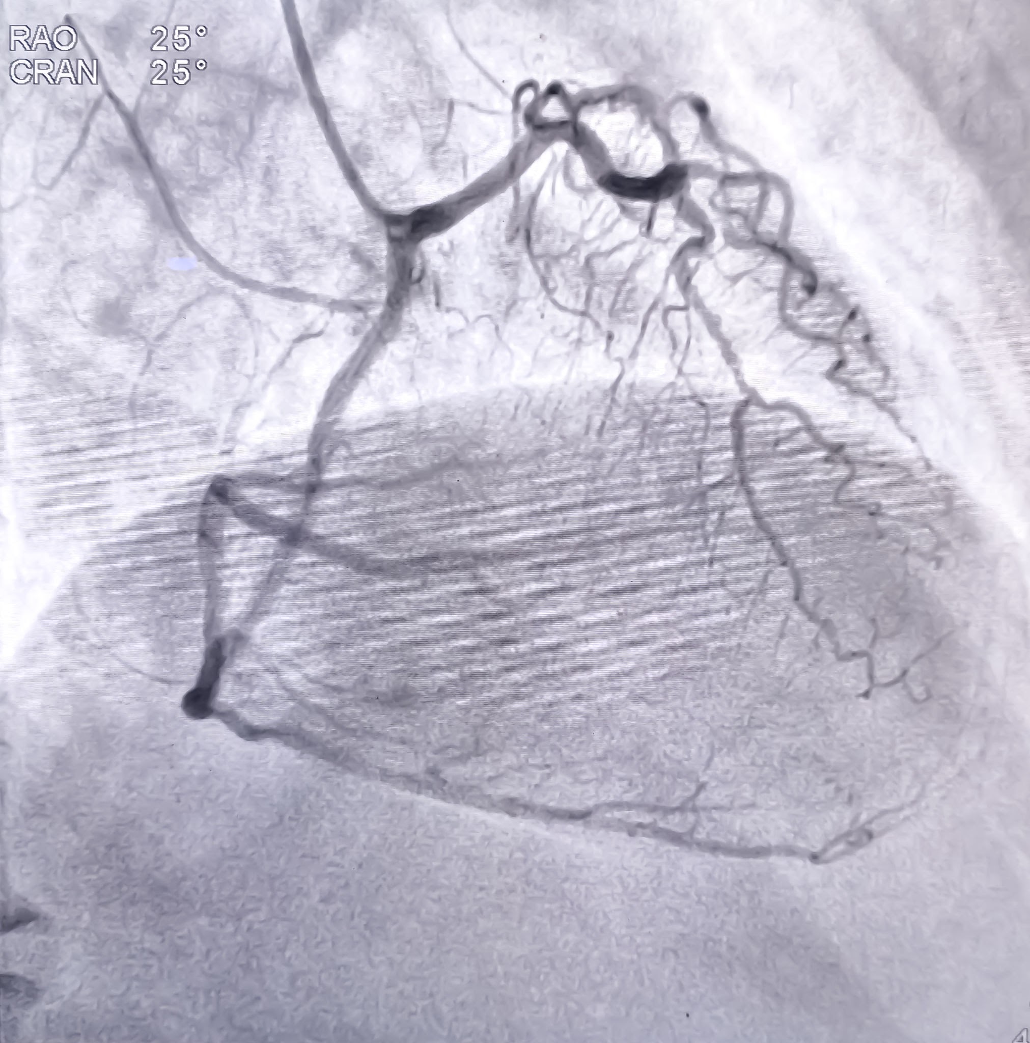
Angiography is a medical imaging procedure used to examine blood vessels and assess how blood flows through the body.
Angiography
What is angiography?
Angiography is a medical imaging procedure used to examine blood vessels and assess how blood flows through the body.
Blood vessels are responsible for carrying oxygen-rich blood from the heart to different organs and returning blood to the heart. When a blood vessel becomes narrowed, blocked, damaged, or abnormally enlarged, it can affect the normal blood supply to surrounding tissues.
During conventional catheter angiography, a special contrast material (dye) is introduced into the blood vessels through a thin tube called a catheter. X-ray images are then taken to make the blood vessels clearly visible.
Angiography helps doctors identify abnormalities such as narrowing, blockages, aneurysms, abnormal blood flow, and certain vascular malformations.
The procedure may also help specialists plan treatment for vascular conditions. In selected cases, treatment can be performed during the same session.
What is catheter angiography?
Catheter angiography is a minimally invasive procedure in which a thin, flexible tube called a catheter is inserted into a blood vessel through a small puncture in the skin.
The catheter is usually introduced through an artery in the wrist or groin, depending on the area being examined and the patient’s condition.
Using continuous X-ray imaging, the specialist carefully guides the catheter through the blood vessels toward the area of interest.
Contrast material is injected through the catheter, making the blood vessels visible on the X-ray images. These images help the specialist evaluate blood flow and identify abnormalities.
Catheter angiography is commonly performed by an interventional radiologist or another specialist trained in vascular procedures.
Why might I need an angiography?
Your doctor may recommend angiography when symptoms, physical examination, or other imaging tests suggest a problem with blood circulation.
Common reasons include:
- Suspected narrowing or blockage of an artery
- Reduced blood circulation in the legs or arms
- Suspected aneurysm or abnormal enlargement of a blood vessel
- Investigation of blood vessel abnormalities in the brain or neck
- Suspected blockage in the blood vessels supplying the kidneys
- Assessment of blood vessels supplying the liver or other abdominal organs
- Investigation of abnormal blood flow or vascular malformations
- Evaluation of certain causes of internal bleeding
- Assessment of blood vessels before a planned surgical or minimally invasive procedure
- Further investigation when non-invasive imaging does not provide sufficient information
The specific reason for angiography depends on your symptoms, medical history, and previous test results.
Which conditions can angiography help diagnose?
Angiography can help investigate several conditions affecting arteries and veins throughout the body.
These may include:
- Peripheral Artery Disease (PAD): Narrowing or blockage of arteries that supply blood to the legs and arms.
- Atherosclerosis: A condition in which fatty deposits build up inside arteries, causing them to narrow.
- Aneurysms: Abnormal enlargement or bulging of a blood vessel.
- Blood vessel blockages: Reduced or interrupted blood flow caused by a clot or other obstruction.
- Carotid artery disease: Narrowing of the arteries in the neck that supply blood to the brain.
- Renal artery disease: Narrowing or other abnormalities affecting the arteries supplying the kidneys.
- Vascular malformations: Abnormal connections or arrangements of blood vessels.
- Internal bleeding: May help identify the blood vessel responsible for bleeding in selected cases.
- Tumour-related vascular abnormalities: May help assess the blood supply to certain tumours.
The findings help doctors understand the location and extent of a vascular problem and decide whether further treatment is required.
What are the different types of angiography?
Angiography can be performed to examine blood vessels in different parts of the body. The type depends on the area being investigated.
- 1. Peripheral Angiography
Used to examine blood vessels in the arms and legs. It may help identify narrowing or blockages associated with reduced circulation. - 2. Cerebral Angiography
Used to examine blood vessels in and around the brain. It may help investigate aneurysms, vascular malformations, and other abnormalities affecting the brain’s circulation. - 3. Carotid Angiography
Used to examine the carotid arteries in the neck, which supply blood to the brain. It may help assess narrowing or blockage in these vessels. - 4. Renal Angiography
Used to examine the arteries supplying the kidneys. It may help identify narrowing, blockages, or other vascular abnormalities. - 5. Visceral Angiography
Used to examine blood vessels supplying abdominal organs such as the liver, spleen, and intestines. - 6. Coronary Angiography
Used to examine the coronary arteries that supply blood to the heart muscle. It is generally performed by an interventional cardiologist.
The appropriate type of angiography depends on the suspected condition and the blood vessels that need to be examined.
How is an angiography performed?
Catheter angiography is generally performed in a specialised angiography suite or interventional radiology department.
The patient lies on an examination table, and the healthcare team monitors vital signs throughout the procedure.
The procedure generally involves the following steps:
- 1. Preparation
The healthcare team reviews the patient’s medical history, medications, allergies, blood test results, and previous imaging. An intravenous (IV) line may be placed in the arm to administer medication or fluids when required. - 2. Preparation of the access site
The selected access area, usually the wrist or groin, is cleaned with an antiseptic solution and covered with sterile drapes. - 3. Local anaesthesia
Local anaesthetic is injected into the skin and surrounding tissues to numb the access area. The patient may feel a brief stinging sensation during the injection.
Sedation may also be provided when appropriate. - 4. Catheter insertion
A small puncture is made in the skin, and a thin catheter is inserted into the selected blood vessel. - 5. Image-guided catheter navigation
Using continuous X-ray imaging, the specialist carefully guides the catheter toward the blood vessels that need to be examined. - 6. Contrast injection and imaging
Contrast material is injected through the catheter. A series of X-ray images are captured as the contrast flows through the blood vessels.
The images help the specialist identify narrowing, blockages, aneurysms, or other vascular abnormalities. - 7. Completion
Once the necessary images have been obtained, the catheter
The patient is then moved to a recovery area for observation.
Is angiography a minimally invasive procedure?
Yes. Catheter angiography is considered minimally invasive because it is performed through a small puncture in the skin rather than a large surgical incision.
The procedure generally uses local anaesthesia, and patients are usually awake during the examination. Sedation may be provided when needed.
X-ray guidance allows the specialist to navigate the catheter through the blood vessels without making a surgical opening along the entire route.
Many patients can return home on the same day after an observation period, although some may require an overnight stay depending on their condition and whether additional treatment was performed.
Who may be a suitable candidate for angiography?
Angiography may be recommended for patients who require detailed examination of their blood vessels.
It may be considered when:
- Symptoms suggest reduced or abnormal blood circulation.
- Ultrasound, CT, or MRI identifies a possible vascular abnormality.
- A suspected blockage or narrowing requires further assessment.
- An aneurysm or vascular malformation needs detailed evaluation.
- The specialist needs more detailed information before planning treatment.
- A vascular condition may require catheter-based treatment.
Not every patient with suspected vascular disease requires catheter angiography. Non-invasive tests such as Doppler ultrasound, CT angiography, or MR angiography may be sufficient in many cases.
Your doctor will assess the expected benefits and risks before recommending the procedure.
Can angiography also be used to treat blocked blood vessels?
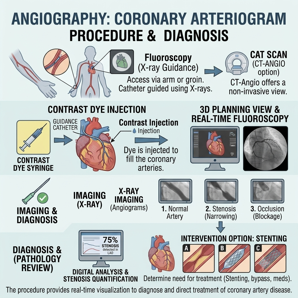
Yes. In selected cases, catheter angiography can be combined with treatment during the same procedure.
Depending on the vascular condition, the specialist may perform:
- Angioplasty: A small balloon is inflated inside a narrowed blood vessel to improve blood flow.
- Stent placement: A small mesh tube is placed inside a blood vessel to help keep it open.
- Embolization: Special materials are introduced into selected blood vessels to block abnormal blood flow or control bleeding.
- Thrombolysis: Medication may be delivered to help dissolve certain blood clots in selected circumstances.
- Other vascular interventions: Additional catheter-based treatments may be considered depending on the location and nature of the abnormality.
Not every angiography procedure includes treatment. The appropriate approach depends on the findings and the patient’s individual condition.
How should I prepare for an angiography?
Preparation depends on the type of angiography, the patient’s medical condition, and whether sedation or additional treatment is planned.
Before the procedure:
- Inform your doctor about all medications you take, including blood thinners, diabetes medication, vitamins, and supplements.
- Tell the healthcare team about any previous allergic reaction to contrast material or other medications.
- Inform your doctor if you have kidney disease, diabetes, or a bleeding disorder.
- Blood tests may be required to assess kidney function and blood-clotting ability.
- Follow the instructions provided regarding eating and drinking. Fasting may be required if sedation is planned.
- Inform your doctor if you are pregnant or think you may be pregnant.
- Wear comfortable clothing and remove jewellery around the procedure area.
- Arrange for someone to accompany you home if sedation is used.
Important: Do not stop prescribed blood thinners, diabetes medication, or other medicines without specific instructions from your treating doctor.
What happens after an angiography?
After the procedure, the catheter is removed and the access site is treated to prevent bleeding.
You will be taken to a recovery area, where the healthcare team monitors your blood pressure, pulse, and overall condition.
Depending on whether the catheter was inserted through the wrist or groin, you may be asked to keep the affected arm or leg still for a period of time.
After the procedure, you may experience:
- Mild pain or tenderness around the puncture site
- Bruising or slight swelling
- A small lump near the access site
- Temporary warmth or flushing following contrast injection
- Mild discomfort around the wrist or groin
These symptoms generally improve over the following days.
Your healthcare team will provide specific instructions before you leave the hospital.
What are the benefits of angiography?
Angiography provides detailed images of blood vessels and helps doctors evaluate abnormalities affecting blood circulation.
Potential benefits include:
- Detailed vascular imaging: Provides clear images of blood vessels and helps identify narrowing, blockages, and other abnormalities.
- Accurate assessment: Helps determine the location and extent of vascular disease.
- Treatment planning: Provides important information when planning vascular surgery or minimally invasive treatment.
- Image-guided treatment: Allows certain catheter-based treatments to be performed during the same session.
- Minimally invasive approach: Usually requires only a small puncture rather than open surgery.
- Targeted examination: Allows the specialist to examine selected blood vessels in different parts of the body.
The benefits depend on the reason for the examination and the patient’s individual medical condition.
Is angiography safe? What are the possible risks?
Angiography is generally considered a safe procedure when performed by an appropriately trained specialist. However, complications can occur.
Possible risks include:
- Bruising, bleeding, or swelling at the catheter insertion site
- Formation of a blood collection (haematoma) near the puncture site
- Infection at the access site
- Injury to the blood vessel
- Formation of a blood clot
- Temporary or, rarely, more significant kidney injury associated with contrast material
- Allergic reaction to the contrast material
- Exposure to a small amount of radiation from X-ray imaging
- Rare complications such as stroke, heart attack, or serious blood vessel damage, depending on the area being examined and the patient’s condition
The risks vary according to the type of angiography, the patient’s overall health, and whether treatment is performed during the same session.
Your specialist will review your medical history and explain any risks that may apply to you.
How long does an angiography take?
The duration depends on the area being examined and the complexity of the procedure.
A diagnostic catheter angiography commonly takes around 30 minutes to 2 hours. More complex procedures or additional treatment may take longer.
Additional time is required for preparation, recovery, and observation after the procedure.
Many patients can return home on the same day if their condition is stable and the treating team considers discharge appropriate.
How long does it take to recover from angiography?
Recovery varies depending on the access site, the type of angiography, and whether additional treatment was performed.
Many patients return home after a few hours of observation.
For a comfortable recovery:
- Rest for the remainder of the day after the procedure.
- Keep the puncture site clean and follow dressing instructions.
- Avoid heavy lifting and strenuous physical activity for the period advised by your doctor.
- Avoid excessive bending or pressure on the affected wrist or leg, depending on the access site.
- Drink fluids as advised by your healthcare team, particularly if contrast material was used and you have no fluid restriction.
- Take prescribed medications as directed.
- Attend recommended follow-up appointments.
Your doctor will advise when you can resume normal activities, work, exercise, and driving.
Seek urgent medical attention if you experience persistent bleeding, rapidly increasing swelling, severe pain, sudden weakness, difficulty speaking, chest pain, or difficulty breathing.
When will I receive my angiography results?
The images obtained during angiography are reviewed by the specialist performing or interpreting the procedure.
The report may describe:
- The location of the blood vessels examined
- Areas of narrowing or blockage
- The degree of reduced blood flow, where assessable
- The presence of aneurysms or vascular abnormalities
- The condition of the examined blood vessels
- Any treatment performed during the procedure
In some cases, the specialist may discuss preliminary findings before you leave the hospital. A detailed report may be prepared afterward and shared with your referring doctor.
Your doctor will explain the results and discuss whether further treatment or monitoring is needed.
What is the difference between angiography, CT angiography, and MR angiography?
All three techniques are used to examine blood vessels, but they use different imaging methods.
Catheter Angiography
- Uses a thin catheter inserted into a blood vessel.
- Uses contrast material and X-ray imaging.
- Provides detailed images of the selected blood vessels.
- Can be combined with certain treatments during the same session.
- Is an invasive procedure with risks associated with catheter insertion and contrast use.
CT Angiography (CTA)
- Uses a CT scanner and contrast material injected into a vein, usually in the arm.
- Produces detailed cross-sectional and three-dimensional images of blood vessels.
- Does not usually require a catheter to be guided through an artery.
- Is commonly used to investigate vascular conditions before deciding whether an intervention is needed.
MR Angiography (MRA)
- Uses magnetic resonance imaging to examine blood vessels.
- May be performed with or without contrast material, depending on the examination.
- Does not use ionising radiation.
- May be suitable for selected patients who require vascular imaging without conventional X-ray angiography.
The most appropriate imaging method depends on the blood vessels being examined, the suspected condition, kidney function, allergies, and the information required by your doctor.
Frequently asked questions about angiography
Is angiography a major surgery?
No. Catheter angiography is a minimally invasive procedure performed through a small puncture in the skin. It generally does not require a large surgical incision.
Will I be awake during angiography?
Most patients remain awake during the procedure. Local anaesthesia is used at the catheter insertion site, and sedation may be provided to help patients relax when appropriate.
Is angiography painful?
The local anaesthetic injection may cause a brief stinging sensation. You may feel some pressure when the catheter is inserted, but the catheter itself should not cause significant pain. A warm sensation may occur when contrast material is injected.
Will I need to stay in the hospital overnight?
Many patients return home on the same day after a period of observation. An overnight stay may be required depending on the patient’s condition, the type of procedure, or any additional treatment performed.
Can I eat before angiography?
You may be asked to avoid eating and drinking for several hours before the procedure, particularly if sedation is planned. Follow the specific instructions provided by your healthcare team.
Is contrast dye used during angiography?
Yes. Conventional catheter angiography generally uses contrast material to make the blood vessels visible on X-ray images. Inform your doctor if you have had a previous reaction to contrast material or have kidney problems.
Can angiography detect blocked arteries?
Yes. Angiography can help identify narrowing or blockages in arteries and assess blood flow through the affected vessels.
Can angiography and angioplasty be performed together?
Yes. If a suitable narrowing is identified and treatment is appropriate, angioplasty may sometimes be performed during the same session. The decision depends on the findings and the patient’s condition.
Does angiography affect kidney function?
The contrast material used during conventional angiography can affect kidney function in some patients, particularly those with existing kidney disease or other risk factors. Your doctor may check kidney function before the procedure and take precautions when needed.
What should I do if the puncture site starts bleeding after angiography?
Apply firm pressure to the site and seek medical assistance immediately if bleeding does not stop. Rapid swelling, severe pain, or a cold or pale limb also requires urgent medical assessment.