A lymph node biopsy is a medical procedure in which a small sample of cells or tissue is collected from a lymph node for laboratory examination.
Lymph Node Biopsy – Image-Guided Lymph Node Biopsy
What is a lymph node biopsy?
A lymph node biopsy is a medical procedure in which a small sample of cells or tissue is collected from a lymph node for laboratory examination.
Lymph nodes are small, bean-shaped structures that form part of the body’s lymphatic and immune systems. They help filter lymph fluid and contain immune cells that help the body fight infections and other diseases.
Lymph nodes are found throughout the body, including the neck, armpits, chest, abdomen, and groin.
Sometimes lymph nodes become enlarged due to infections, inflammation, immune system conditions, or cancer. A biopsy allows doctors to examine the cells or tissue and investigate the reason for the enlargement.
What is an image-guided lymph node biopsy?
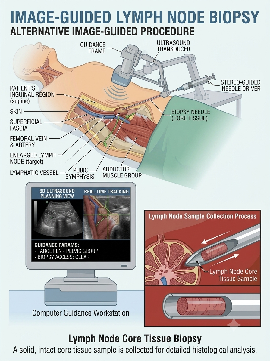
An image-guided lymph node biopsy is a minimally invasive procedure in which ultrasound or CT imaging helps the specialist locate a lymph node and guide a needle into the appropriate area.
A small sample of cells or tissue is collected using a specialised needle and sent to a laboratory for examination.
Image guidance helps the specialist identify the target and plan a safe route for collecting the sample.
Why might I need a lymph node biopsy?
Your doctor may recommend a lymph node biopsy when a lymph node remains enlarged, has an unusual appearance on imaging, or requires further investigation.
Common reasons include:
- Persistent or unexplained enlargement of lymph nodes
- Lymph nodes with unusual features on ultrasound, CT, or MRI
- Suspected lymphoma or another type of cancer
- Investigation of possible cancer spread from another part of the body
- Unexplained swelling in the neck, armpit, chest, abdomen, or groin
- Suspected infections affecting the lymph nodes
- Investigation of certain inflammatory or immune system conditions
- Further evaluation when blood tests and imaging do not provide a clear diagnosis
- Monitoring selected patients with a previously diagnosed condition
Not every enlarged lymph node requires a biopsy. Your doctor will consider its size, appearance, location, duration of enlargement, symptoms, and medical history before recommending the procedure.
What causes enlarged lymph nodes?
Lymph nodes can become enlarged when the immune system responds to an infection, inflammation, or another medical condition.
Common causes include:
- Infections: Viral or bacterial infections can cause nearby lymph nodes to swell.
- Tuberculosis: Tuberculosis can affect lymph nodes, particularly those in the neck and other areas of the body.
- Inflammatory conditions: Certain inflammatory and autoimmune disorders can cause lymph node enlargement.
- Lymphoma: A type of cancer that begins in the lymphatic system.
- Metastatic cancer: Cancer that has spread from another part of the body to the lymph nodes.
- Other conditions: Certain medications and less common medical conditions may also cause lymph node enlargement.
An enlarged lymph node does not automatically mean cancer is present. A biopsy may be recommended when further investigation is needed to identify the cause.
Which conditions can a lymph node biopsy help diagnose?
A lymph node biopsy can help investigate several conditions affecting the lymphatic and immune systems.
These may include:
- Lymphoma: A group of cancers that develop in lymphocytes, which are a type of white blood cell.
- Metastatic cancer: Cancer that has spread from another organ or tissue to the lymph nodes.
- Tuberculosis: An infection that can affect lymph nodes.
- Other infections: Certain bacterial, viral, fungal, and other infections.
- Sarcoidosis: An inflammatory condition that can cause clusters of immune cells to form in different organs, including lymph nodes.
- Other inflammatory or immune system disorders: Conditions that may cause abnormal lymph node changes.
The biopsy findings are interpreted alongside the patient’s symptoms, blood tests, imaging, and other relevant investigations.
What are the different types of lymph node biopsy?
The appropriate biopsy technique depends on the location of the lymph node, the suspected condition, and the amount and type of tissue needed.
The main types include:
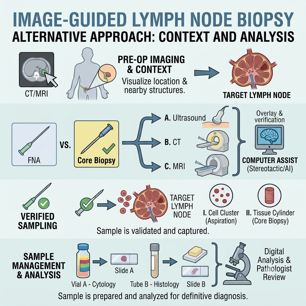
- 1. Fine Needle Aspiration (FNA)
Fine needle aspiration uses a very thin needle attached to a syringe to collect cells or fluid from a lymph node.
It is a relatively quick procedure and may help investigate infections, metastatic cancer, and other abnormalities.
However, FNA collects individual cells rather than a larger piece of tissue. It may not provide enough information to diagnose or classify certain conditions, particularly lymphoma. - 2. Core Needle Biopsy
A core needle biopsy uses a slightly larger hollow needle to collect small pieces of tissue from the lymph node.
It provides tissue samples that preserve more of the lymph node’s structure than FNA.
This can help pathologists examine the arrangement of cells and perform additional tests when required.
Ultrasound or CT guidance may be used to position the needle accurately. - 3. Excisional Biopsy
An excisional biopsy involves surgically removing an entire lymph node for laboratory examination.
It may be recommended when a larger tissue sample is needed, particularly when lymphoma is suspected and the lymph node’s overall structure is important for diagnosis.
This procedure is generally performed by a surgeon and may require local or general anaesthesia depending on the location and complexity. - 4. Incisional Biopsy
An incisional biopsy involves surgically removing only part of a lymph node.
It may be considered when removing the entire lymph node is not practical or appropriate.
Your doctor will recommend the technique that is most suitable for your condition and diagnostic requirements.
How is an ultrasound-guided lymph node biopsy performed?
An ultrasound-guided lymph node biopsy is generally performed in a specialised imaging or interventional radiology department.
The patient is positioned according to the location of the lymph node. For example, a neck biopsy may be performed while lying on the back, whereas an armpit or groin biopsy may require a different position.
The procedure generally involves the following steps:
- 1. Preparation
The healthcare team reviews the patient’s medical history, medications, allergies, and relevant imaging. The patient is positioned comfortably, and the biopsy area is cleaned with an antiseptic solution. - 2. Ultrasound examination
An ultrasound probe is placed over the skin to identify the enlarged lymph node and determine the safest route for needle placement. - 3. Local anaesthesia
Local anaesthetic is injected into the skin and surrounding tissues to numb the biopsy area.
The patient may feel a brief stinging sensation when the anaesthetic is administered. - 4. Needle insertion
A small puncture is made in the skin, and the biopsy needle is guided into the lymph node using real-time ultrasound imaging. - 5. Sample collection
The specialist collects one or more samples depending on the type of biopsy and the amount of tissue required.
Several samples may be needed to provide sufficient material for laboratory examination and additional tests. - 6. Completion
The needle is removed, and pressure is applied to the biopsy site to reduce bleeding. A small dressing is placed over the puncture.
The samples are sent to the laboratory for examination.
Is a lymph node biopsy a minimally invasive procedure?
An image-guided needle biopsy is considered minimally invasive because it is performed through a small puncture in the skin rather than a surgical incision.
It is generally performed using local anaesthesia and may not require sedation.
Ultrasound or CT imaging helps the specialist locate the lymph node and guide the needle toward the selected area.
Many patients can return home on the same day after the procedure.
However, not all lymph node biopsies are needle procedures. An excisional or incisional biopsy requires a surgical approach and may involve different anaesthesia and recovery arrangements.
Who may be a suitable candidate for an image-guided lymph node biopsy?
An image-guided lymph node biopsy may be considered for patients who have an enlarged or abnormal lymph node that requires further investigation.
It may be recommended when:
- A lymph node remains enlarged without an identified cause.
- Imaging shows features that require tissue examination.
- There is a suspected infection or inflammatory condition.
- A doctor needs to investigate possible cancer involvement.
- A lymph node is located in an area that can be safely accessed using imaging guidance.
- A tissue sample is needed to guide further diagnosis or treatment.
The suitability of a needle biopsy depends on the location of the lymph node, the suspected condition, and the amount of tissue required.
If lymphoma is suspected, a core needle or surgical excisional biopsy may be preferred in some situations because the structure of the lymph node is important for diagnosis.
Your specialist will determine the most appropriate method after reviewing your clinical findings and imaging.
How long does a lymph node biopsy take?
The duration depends on the location of the lymph node and the type of biopsy performed.
An ultrasound-guided needle biopsy commonly takes around 15–30 minutes, although additional time may be required for preparation, sample collection, and observation.
A CT-guided biopsy or surgical biopsy may take longer depending on the complexity of the procedure.
Many patients undergoing a needle biopsy can return home on the same day if recovery is satisfactory.
Is a lymph node biopsy painful?
Most patients tolerate an image-guided needle biopsy well.
Local anaesthesia is used to numb the skin and surrounding tissues before the needle is inserted.
You may experience:
- A brief stinging sensation when local anaesthetic is injected
- Mild pressure when the biopsy needle is positioned
- Temporary discomfort during sample collection
- Mild soreness or tenderness after the procedure
If you experience significant pain or discomfort, inform the specialist or healthcare team.
How should I prepare for a lymph node biopsy?
Preparation depends on the location of the lymph node and the type of biopsy being performed.
Before your appointment:
- Inform your doctor about all medications you take, including blood thinners, aspirin, vitamins, and supplements.
- Tell the healthcare team about any medication allergies.
- Inform your doctor if you have a bleeding disorder or a history of unusual bleeding.
- Follow instructions regarding eating and drinking. Fasting may not be necessary for a needle biopsy performed with local anaesthesia alone.
- Wear comfortable clothing that allows easy access to the biopsy area.
- Ask whether you need someone to accompany you home if sedation is planned.
Important: Do not stop blood thinners or other prescribed medications without specific instructions from your treating doctor.
What happens after a lymph node biopsy?
After the sample is collected, the needle is removed, and pressure is applied to the biopsy site to reduce bleeding.
A small dressing is placed over the puncture, and the healthcare team may observe you briefly before discharge.
After the procedure, you may experience:
- Mild pain or tenderness at the biopsy site
- Slight bruising or swelling
- Temporary discomfort when moving the affected area
- Minor bleeding or spotting around the dressing
These symptoms generally improve over the following days.
Follow the wound-care instructions provided by your healthcare team.
What are the benefits of an image-guided lymph node biopsy?
An image-guided lymph node biopsy provides tissue or cell samples that can help doctors investigate the cause of an abnormal lymph node.
Potential benefits include:
- Minimally invasive approach: Needle biopsy requires only a small puncture in the skin.
- Image guidance: Ultrasound or CT helps identify the lymph node and guide needle placement.
- Diagnostic information: Helps investigate infections, inflammatory conditions, and cancer.
- Targeted sampling: Allows the specialist to collect samples from a specific lymph node.
- Outpatient procedure: Many needle biopsies do not require an overnight hospital stay.
- Treatment planning: Laboratory findings can help doctors decide whether further testing or treatment is required.
The diagnostic value depends on the type and adequacy of the sample collected.
Is a lymph node biopsy safe? What are the possible risks?
Image-guided lymph node needle biopsy is generally considered safe. However, complications can occur.
Possible risks include:
- Pain or tenderness around the biopsy site
- Bruising or swelling
- Minor bleeding or a collection of blood beneath the skin
- Infection at the puncture site
- Injury to nearby blood vessels or structures
- Temporary numbness or discomfort, depending on the biopsy location
- An inadequate sample that may require repeat biopsy or another procedure
The risks vary depending on the location of the lymph node and the biopsy technique.
Your specialist will review your medical history and imaging findings before the procedure and explain any risks that may apply to you.
How long does it take to recover from a lymph node biopsy?
Recovery after an image-guided needle biopsy is usually quick.
Many patients can resume light daily activities on the same day or the following day, depending on how they feel and the location of the biopsy.
Mild soreness or bruising may last for several days.
For a comfortable recovery:
- Keep the dressing in place for the period advised by your healthcare team.
- Follow instructions for keeping the biopsy site clean and dry.
- Avoid heavy lifting or strenuous activity for the period recommended.
- Avoid unnecessary pressure on the biopsy area.
- Take pain relief only as recommended by your doctor.
- Attend follow-up appointments as advised.
Contact your healthcare provider if you notice increasing pain, swelling, redness, discharge, fever, or persistent bleeding.
When will I receive my lymph node biopsy results?
The collected samples are sent to a laboratory for examination by a pathologist.
Depending on the suspected condition, the sample may undergo several types of testing, including:
- Microscopic examination: Evaluates the appearance and arrangement of cells and tissue.
- Immunohistochemistry: Uses special markers to help identify the type of cells present.
- Flow cytometry: Helps identify abnormal populations of immune cells and is often useful when lymphoma is suspected.
- Microbiological testing: May be performed when an infection such as tuberculosis is suspected.
- Molecular or genetic testing: May be required to investigate specific diseases or classify certain cancers.
Not every patient requires all these tests.
Results may take several days or longer, depending on the type of testing required. Your treating doctor will explain the findings and discuss the next steps.
Can a lymph node biopsy detect cancer?
Yes. A lymph node biopsy can help identify cancer cells within a lymph node.
It may help diagnose:
- Lymphoma, which begins in the lymphatic system
- Metastatic cancer that has spread from another organ
- Other abnormal growths affecting the lymph nodes
The biopsy can also help identify the type of abnormal cells and guide additional testing.
However, an abnormal or enlarged lymph node does not automatically mean cancer is present. Infections and inflammatory conditions can also cause lymph node enlargement.
Can a needle biopsy diagnose lymphoma?
A needle biopsy can sometimes help diagnose lymphoma, but the amount and type of tissue collected are important.
Fine needle aspiration collects individual cells and may not provide enough information to establish or classify lymphoma.
Core needle biopsy provides a larger tissue sample and may be useful in selected cases. However, an excisional biopsy that removes the entire lymph node may be recommended when the pathologist needs to examine the overall structure of the lymph node.
The appropriate method depends on the suspected type of lymphoma, the location of the lymph node, and the clinical circumstances.
What happens if the lymph node biopsy results are inconclusive?
Sometimes the biopsy sample does not provide enough information to establish a clear diagnosis.
This may happen if the sample contains too few cells, does not include enough tissue, or does not preserve the structure needed for diagnosis.
Depending on the findings, your doctor may recommend:
- A repeat needle biopsy
- A core needle biopsy if an earlier FNA was insufficient
- A surgical excisional biopsy
- Additional laboratory testing
- Further imaging
- Consultation with a specialist, such as a haematologist or oncologist
The next step depends on the suspected condition and the results of the initial biopsy.
What is the difference between a lymph node biopsy and a lymph node aspiration?
Both procedures collect samples from a lymph node, but they obtain different types of material.
Lymph node aspiration (FNA)
- Uses a very thin needle to collect cells or fluid.
- Usually requires only a small puncture in the skin.
- Can help investigate certain infections and metastatic cancers.
- May not provide enough tissue for diagnosing or classifying some lymphomas.
Lymph node core biopsy
- Uses a larger hollow needle to collect small pieces of tissue.
- Preserves more of the tissue structure.
- Can provide additional information for diagnosing certain lymph node conditions.
- May be preferred when a larger tissue sample is needed.
Your doctor will recommend the appropriate method based on the diagnostic question.
Frequently asked questions about lymph node biopsy
Is a lymph node biopsy a major surgery?
An image-guided needle biopsy is not major surgery. It is generally performed through a small puncture in the skin using local anaesthesia. A surgical excisional or incisional biopsy is a different procedure and involves a small surgical incision.
Will I need to stay in the hospital overnight?
Most patients undergoing an image-guided needle biopsy can return home on the same day after a short observation period. Surgical biopsies may require different arrangements depending on the anaesthesia and procedure.
Can I eat before a lymph node biopsy?
Fasting is often unnecessary for a needle biopsy performed with local anaesthesia alone. However, different instructions may apply if sedation or general anaesthesia is planned. Follow the instructions provided by your healthcare team.
Can I take my regular medicines before the biopsy?
Some medications, particularly blood thinners, may require special instructions before the procedure. Do not stop prescribed medications without consulting your doctor.
Will I have a scar after a lymph node biopsy?
A needle biopsy generally leaves a very small puncture mark that usually heals with minimal visible scarring. A surgical biopsy may leave a small incision scar.
Can an enlarged lymph node be harmless?
Yes. Many enlarged lymph nodes occur because of infections or inflammation and are not cancerous. A biopsy may be recommended when the cause remains uncertain or further investigation is necessary.
How long will the biopsy site remain sore?
Mild soreness or tenderness may last for several days. Contact your healthcare provider if the discomfort becomes severe or continues to worsen.
Can a lymph node biopsy be repeated?
Yes. A repeat biopsy may be recommended if the first sample is insufficient, the results are inconclusive, or additional tissue is needed for further testing.
What should I do if I notice swelling or bleeding after the biopsy?
Minor bruising or tenderness can occur. However, increasing swelling, persistent bleeding, severe pain, fever, or discharge should be reported to your healthcare provider.
Need further evaluation of an enlarged lymph node?
An image-guided lymph node biopsy can help investigate unexplained swelling and provide important information about infections, inflammatory conditions, and possible cancer.